


Myosin proteins form a large superfamily of ATP-dependent motor proteins essential for muscle contraction, cytokinesis, cell migration, intracellular transport, and organelle positioning. Dysregulation of myosin activity is linked to numerous diseases, including cardiomyopathies, skeletal muscle disorders and cancer, making myosin isoforms highly attractive therapeutic targets [1]. With the clinical success of selective cardiac myosin inhibitors, such as mavacamten, for obstructive hypertrophic cardiomyopathy, interest in myosin-targeted drug discovery has rapidly expanded [2].
To contribute to this growing field of myosin-targeted drug discovery, Life Chemicals has developed two complementary screening collections using advanced ligand-based and structure-based approaches:
- Myosin Focused Library (1,200 compounds in stock)
- Myosin Targeted Library (2,000 compounds, synthesized on request)
Together, they provide drug-like small molecules optimized for high-throughput screening (HTS) and hit-to-lead optimization across multiple myosin isoforms.
 The compound selection can be customized based on your requirements. Cherry-picking is available.
 Please contact us at orders@lifechemicals.com for any additional information and price quotations.

Further exploring our related products will make your search even more rewarding:
Representative screening compounds from the Myosin-focused Screening Library


Myosin Focused Library
This ligand-based screening set comprises approximately 1,200 drug-like compounds in stock that exhibit potential activity against diverse myosin proteins and are well-suited for primary screening and early SAR exploration in myosin-driven projects.
First, a reference set of experimentally validated myosin inhibitors was extracted exclusively from the ChEMBL database (IC₅₀, Kᵢ, or related activity values < 10 μM; inhibition > 25%). These reference compounds were then used to perform 2D fingerprint similarity screening against the proprietary HTS Compound Collection using a Tanimoto coefficient ≥ 0.75. This approach yielded a focused, chemically diverse screening library enriched with compounds potentially active against multiple myosin isoforms, including:
- Myosin-IIIa
- Myosin-IIIb
- Myosin light chain kinase 2 (skeletal/cardiac muscle)
- Myosin light chain kinase family member 4
- Smooth muscle myosin light chain kinase

Figure 1. Compound distribution targeting protein within the Myosin Focused Library.
![Figure 2. Cardiac Myosin Inhibitors in hypertrophic cardiomyopathy [3].](img/library_descriptions/Myosin-focused_Screening_Library/Myosin-focused_Screening_Library_2.png)
Figure 2. Cardiac Myosin Inhibitors in hypertrophic cardiomyopathy [3].
Myosin Targeted Library
This structure-based docking set comprises over 2,000 drug-like screening compounds, synthesizable on request, designed to selectively interact with key human myosin targets, including:
It has been developed using structure-guided combinatorial chemistry, guided by the binding modes and pharmacophoric features of known myosin inhibitors, particularly mavacamten (a selective inhibitor of β-cardiac myosin) and sevasemten (a myosin-2 inhibitor). Virtual combinatorial libraries were generated based on these core scaffolds and prioritized by molecular docking into the validated ligand-binding sites of the target myosin isoforms.
The selected optimized small-molecule analogs exhibited improved predicted binding affinity and interaction profiles compared to the reference inhibitors, indicating enhanced inhibitory potential and strong suitability for hit identification and lead optimization.
Myosin-2
Myosin-2 is a major contractile protein in eukaryotic cells that converts ATP hydrolysis into mechanical force, enabling movement along actin filaments [4]. It is essential for muscle contraction and also participates in key cellular processes such as cell division, adhesion, and motility [4-5]. Structurally, myosin-2 consists of two heavy chains, two essential light chains (ELCs), and two regulatory light chains (RLCs) [4]. The heavy chains form a globular head with actin-binding and ATPase activity and an α-helical rod that assembles into a coiled-coil tail capable of filament formation [4-5]. The ATPase activity of myosin-2 is precisely regulated: in its phosphorylated state, it forms filaments and hydrolyzes ATP. In contrast, in the dephosphorylated state it adopts a compact, kinetically inert conformation (>1000-fold reduced ATPase activity), also known as the interacting heads motif (IHM) [4].
Myosin-2 is a potential therapeutic target in conditions associated with excessive muscle contraction. Direct inhibition of skeletal myosin-2 may be used to treat spasticity and muscle stiffness following nervous system injuries (e.g., brain or spinal cord injury, stroke, cerebral palsy), as well as disorders involving upper motor neuron lesions, certain myopathies, and conditions such as low back pain and fibromyalgia [6].
Molecular docking of the combinatorial library was performed into the binding sites on the surface of human Myosin-2, using the AlphaFold-predicted structure. Two putative surface sites were identified (Figure 3): site 5, corresponding to ADP binding, and site 2, corresponding to the binding of potential inhibitors.
Docking was also carried out in the binding site of the selective inhibitor MPH-220 (PDB ID: 6YSY [6]) (Figure 4).

Figure 3. Prediction of potential binding sites on the surface of human myosin-2.

Figure 4. ADP and selective inhibitor MPH-220 binding sites (PDB ID: 6YSY).
The predicted site 2 partially overlaps with the MPH-220 binding site; however, the predicted site displays a larger and slightly shifted surface. As a result, 554 compounds were selected as hits for the MPH-220 binding site, with docking scores ≤ –6. The dataset was further complemented with the best unique hits for site 2. Compounds binding to site 5 were not considered due to the low probability of competitive inhibition.
Key features:
- Method: SP (standard precision) ligand-receptor docking
- X-Ray/3D data used: AlphaFold, 6YSY
- Constraints: no
- Filters used: no
- Number of compounds selected: 908
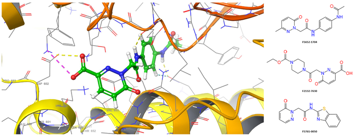
Figure 5. Spatial structure binding site of the complex of myosin-2 with the lead docking molecule F2132-7571 (docking score = -7.46).
Myosin-7 (beta-cardiac myosin)
β-cardiac myosin is the molecular motor responsible for force generation in cardiac ventricular tissue, driven by ATP hydrolysis and actin filament binding. It consists of two heavy chains and four light chains (two essential and two regulatory); each heavy chain contains an N-terminal motor domain and an α-helical region forming a coiled coil. Myosin molecules form bipolar filaments and generate force through cyclic interactions with actin during the ATPase cycle [7].
Hypertrophic cardiomyopathy (HCM) is a genetic disease of the sarcomere, with ~40 % of mutations located in the β-cardiac myosin heavy chain gene (MYH7), making it a key disease target [7]. HCM is associated with increased availability of myosin heads for actin interaction and force production, leading to impaired relaxation. In contrast, some mutations causing dilated cardiomyopathy stabilize the autoinhibited state, thereby reducing force production [7-8]. β-cardiac myosin is therefore targeted by small-molecule modulators such as Mavacamten, which inhibit ATPase activity, stabilize the autoinhibited interacting-heads motif and reduce contractile force, representing a therapeutic approach to disease progression [7].
Key features:
- Method: SP (standard precision) ligand-receptor docking
- X-Ray data used: 9GZ2
- Constraints: no
- Filters used: no
- Number of compounds selected: 1,114
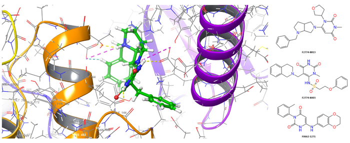
Figure 6. Spatial structure of the binding site of the complex of beta-cardiac myosin with the lead docking molecule F2774-8044 (docking score = -10.16).
Reference:
- Ostrominski, J, Guo, R, Elliott, P. et al. Cardiac Myosin Inhibitors for Managing Obstructive Hypertrophic Cardiomyopathy: JACC: Heart Failure State-of-the-Art Review. J Am Coll Cardiol HF. 2023 Jul, 11 (7) 735–748. https://doi.org/10.1016/j.jchf.2023.04.018
- Ramadan MM, Al-Najjar RA, Abady RS, Obaid HA, Mostafa YA, Al-Obeid MT, Elmahal M. Mavacamten Cardiac Myosin Inhibitor: Clinical Applications and Future Perspectives. Cureus. 2025 Apr 21;17(4):e82722. doi: 10.7759/cureus.82722. PMID: 40400802; PMCID: PMC12094493.
- Hou L, Lin B, Ji X, Huang A. Cardiac Myosin Inhibitors in Hypertrophic Cardiomyopathy: Clinical Advances and Therapeutic Prospects. Drug Des Devel Ther. 2025 Oct 17;19:9367-9385. doi: 10.2147/DDDT.S555335. PMID: 41127363; PMCID: PMC12539420.
- Heissler SM, Arora AS, Billington N, Sellers JR, Chinthalapudi K. Cryo-EM structure of the autoinhibited state of myosin-2. Sci Adv. 2021 Dec 24;7(52):eabk3273. doi: 10.1126/sciadv.abk3273. Epub 2021 Dec 22. PMID: 34936462; PMCID: PMC8694606.
- Bharda AV, Jung HS. A simple and rapid preparation of smooth muscle myosin 2 for the electron microscopic analysis. Appl Microsc. 2024 Jan 2;54(1):1. doi: 10.1186/s42649-023-00094-5. PMID: 38165512; PMCID: PMC10761634.
- Gyimesi M, Horváth ÁI, Túrós D, et al. Single Residue Variation in Skeletal Muscle Myosin Enables Direct and Selective Drug Targeting for Spasticity and Muscle Stiffness. Cell. 2020;183(2):335-346.e13. doi:10.1016/j.cell.2020.08.050
- McMillan SN, Pitts JRT, Barua B, Winkelmann DA, Scarff CA. Mavacamten inhibits myosin activity by stabilizing the myosin-interacting heads motif and stalling motor force generation. bioRxiv [Preprint]. 2025 Feb 17:2025.02.12.637875. doi: 10.1101/2025.02.12.637875. PMID: 39990378; PMCID: PMC11844505.
- Goluguri RR, Guhathakurta P, Nandwani N, Dawood A, Yokota S, Roopnarine O, Thomas DD, Ruppel KM, Spudich JA. A FRET assay to monitor different structural states of human β-cardiac myosin including the interacting-heads motif. Proc Natl Acad Sci U S A. 2025 Aug 26;122(34):e2504562122. doi: 10.1073/pnas.2504562122. Epub 2025 Aug 20. PMID: 40833405; PMCID: PMC12403093.